Hematocrit and Hemoglobin: 7 Critical Differences
Hematocrit and Hemoglobin: 7 Critical Differences
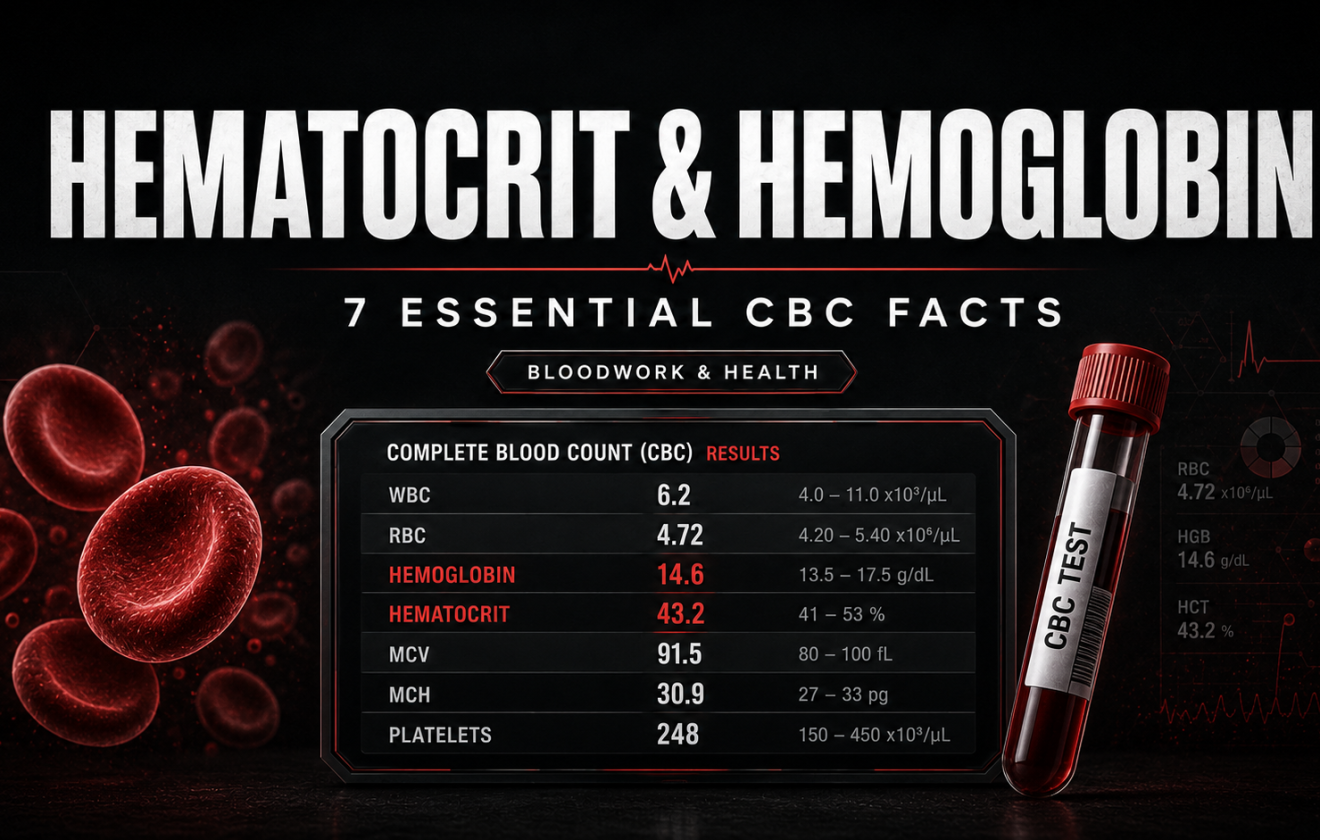
Hematocrit and hemoglobin are two different readings taken from one tube of blood. Hemoglobin is a concentration, the grams of oxygen carrying protein in every decilitre. Hematocrit is a proportion, the share of blood volume occupied by packed red cells. Hematocrit and hemoglobin usually rise and fall together, they come apart in the situations that matter most to lifters, and on testosterone it is hematocrit rather than hemoglobin that decides whether a dose continues. Dose management itself lives on high hematocrit on TRT.
Hematocrit vs Hemoglobin: The Data in Brief
Hemoglobin measures oxygen carrying protein per decilitre of blood. Hematocrit measures what fraction of that volume is red cells. Hematocrit and hemoglobin are therefore a proportion and a concentration, which is why they are not interchangeable even though a laboratory prints them side by side.
In a randomised trial of testosterone gel in older men, hemoglobin rose about 7 percent while hematocrit rose about 10 percent, with erythropoietin up and hepcidin and ferritin down (Bachman E et al, J Gerontol A Biol Sci Med Sci, 2014, PMID 24158761). Hematocrit and hemoglobin do not move by the same amount.
Of hematocrit and hemoglobin, only the first carries a dosing rule. The Endocrine Society advises against starting testosterone at a baseline hematocrit above 48 percent and recommends dose reduction or interruption above 54 percent (Bhasin S et al, J Clin Endocrinol Metab, 2018, PMID 29562364).
Every number quoted here about hematocrit and hemoglobin is traceable to a named publication and its PubMed identifier, so a claim and its source can be checked together rather than taken on trust.
What This CBC Guide Covers
The comparison itself. What hematocrit and hemoglobin each measure, the units they use, reference ranges by sex, what raises and lowers each, and which one a clinician acts on.
Panel context. Where hematocrit and hemoglobin sit inside a complete blood count, how the red cell indices relate to them, and when the usual arithmetic between the pair breaks down.
The trained body. Plasma volume expansion, dehydration, draw timing, altitude and exercise hemolysis, and why hematocrit and hemoglobin read differently in a trained man.
Androgen physiology. Why testosterone drives erythrocytosis, how large the effect on hematocrit and hemoglobin is, and why only one of them is written into dosing rules.
No protocols. No doses, no cycle design and no instructions for lowering a reading at home appear anywhere on this page.
Management on therapy. Dose adjustment, donation intervals and monitoring schedules belong to high hematocrit on TRT.
Compound specific effects. Which anabolic agents push red cell mass hardest is covered on do steroids cause high hematocrit.
Arithmetic. Converting a reading into a zone is handled by the hematocrit calculator.
Hematocrit vs Hemoglobin: What Each One Measures
The short answer to hematocrit vs hemoglobin is this. Hemoglobin is a concentration, hematocrit is a proportion. A hemoglobin of 15.2 g/dL says every decilitre of whole blood holds 15.2 grams of the iron bearing protein that binds oxygen. A hematocrit of 45 percent says that if the same blood were spun down, packed red cells would fill 45 percent of the column. One counts cargo, the other counts space.
Because red cells are the only place hemoglobin lives, hematocrit and hemoglobin are tightly coupled in healthy adults. Analysers exploit that coupling. Most instruments measure hemoglobin directly, count and size red cells electronically, then calculate hematocrit as red cell count multiplied by mean corpuscular volume. On many reports the hematocrit was never measured at all, which is the first practical difference between hematocrit and hemoglobin worth knowing.
That coupling produces a sanity check laboratory staff still use. In a person with normally sized red cells, hematocrit is close to three times hemoglobin. A hemoglobin of 15 g/dL pairs with a hematocrit near 45 percent, 16 with 48, 13 with 39. When hematocrit and hemoglobin drift away from that ratio, the cells have changed size, and the mismatch is information rather than an error.
That is where the second difference bites. Iron deficiency shrinks red cells, so hemoglobin can fall faster than hematocrit. B12 or folate deficiency enlarges them, so hematocrit can hold up while hemoglobin sags. Plasma volume shifts move hematocrit hard while leaving total body hemoglobin untouched. One aside for readers whose laboratory prints the other spelling: haematocrit and haemoglobin are the same pair of measurements, and nothing in the physiology changes with the extra letter.
| Property | Hematocrit | Hemoglobin | Status |
|---|---|---|---|
| What it measures | Percentage of blood volume occupied by packed red cells | Grams of oxygen carrying protein per decilitre of whole blood | ● Acted on |
| Units | Percent, or a decimal fraction such as 0.45 | Grams per decilitre, or grams per litre in Europe | ● Background |
| How the analyser gets it | Usually calculated from cell count times cell volume | Measured directly by a chemical method | ● Watched |
| Typical adult male range | 40 to 52 percent, with 52 to 54 treated as borderline | 13.5 to 17.5 g/dL, with 17.5 to 18.5 treated as borderline | ● Acted on |
| Typical adult female range | 36 to 48 percent, with 48 to 50 treated as borderline | 12.0 to 15.5 g/dL, with 15.5 to 16.5 treated as borderline | ● Acted on |
| What raises it | Androgens, altitude, smoking, sleep apnoea, dehydration, diuretics, polycythaemia vera | The same drivers, minus most of the plasma effects | ● Acted on |
| What lowers it | Bleeding, haemolysis, iron deficiency, chronic disease, plasma expansion, pregnancy | The same list, but plasma expansion barely changes total body content | ● Watched |
| Sensitivity to hydration | High. A dehydrated draw inflates it | Lower in practice, though the same sample effect applies | ● Acted on |
| Which one guidelines act on | Carries the 54 percent testosterone threshold | Defines anaemia and judges oxygen carrying capacity | ● Acted on |
| What it tells you about symptoms | Viscosity and clotting risk at the high end | Fatigue and breathlessness at the low end | ● Watched |
Read the last two rows upward and the division of labour between hematocrit and hemoglobin is clear. Hemoglobin is the anaemia marker, answering whether enough oxygen carrying protein is present. Hematocrit is the viscosity marker, answering how thick the blood has become. Most decisions that affect men on hormones live at the thick end, which is why hematocrit carries the thresholds.
Where Hematocrit and Hemoglobin Sit Inside a CBC
A complete blood count reports white cells, platelets and a red cell block. Inside that block, hematocrit and hemoglobin sit beside the red cell count, the indices and the distribution width, and reading them together is what separates interpretation from guesswork. The baseline panel guide shows how the panel fits a baseline workup.
Red cell count is the number of cells per litre. Mean corpuscular volume is average cell size. Mean corpuscular hemoglobin is the average protein mass inside one cell. Red cell distribution width describes how variable the sizes are. Hematocrit and hemoglobin are the two summary figures that these indices exist to explain.
So an abnormal result is rarely interpretable alone. Low hemoglobin with a low mean corpuscular volume and a wide distribution width points at iron deficiency. Low hemoglobin with a high mean corpuscular volume points at B12, folate or alcohol. A high hematocrit with normal indices, normal white cells and normal platelets points at plasma volume or a secondary drive such as androgen exposure, not at marrow disease.
Where the lower limit of normal sits is less fixed than a report implies. Reference intervals for hematocrit and hemoglobin come from the distribution of values in a reference population, and the percentile chosen as abnormal is a statistical decision rather than a biological boundary (Beutler E, Waalen J, Blood, 2006;107(5):1747-50, PMID 16189263). A value one point below a printed limit is not a diagnosis.
Hematocrit is often derived, not measured. If mean corpuscular volume is wrong, because the sample sat too long and the cells swelled, the calculated hematocrit inherits that error while the directly measured hemoglobin does not.
Units differ by country. Hematocrit may appear as 45 percent or as 0.45, and hemoglobin as 15.2 g/dL or 152 g/L. Comparing a result from one laboratory with a threshold from another without checking units is a common and avoidable error.
A single reading is a snapshot. Hematocrit and hemoglobin both move with posture, time of day, hydration and recent training. A trend across three draws taken under similar conditions beats one alarming value.
Hematocrit and Hemoglobin: Reference Bands and Event Rates
The chart sets hematocrit and hemoglobin side by side on their own scales, typical adult reference band in green, elevated zone in red. The asymmetry is obvious. The hemoglobin scale is narrow and the hematocrit scale is wide, so the same physiological push registers as a larger looking move on the right hand panel.

The animation below shows different data. Not the reference bands for hematocrit and hemoglobin, but how often drug induced erythrocytosis actually occurs. A systematic review of 45 studies found erythrocytosis rates of up to 66.7 percent in cisgender and transgender men on prescription testosterone, with intramuscular formulations, higher doses and older age carrying the greater risk, and up to 2.7 percent of men on testosterone therapy developing a thromboembolic event. Rates on SGLT-2 inhibitors ranged from 2.1 to 22 percent (Liu J et al, Blood Adv, 2025;9(9):2108-2118, PMID 39913688).
Reported rates of drug induced erythrocytosis and of thromboembolic events, plotted on a common 0 to 70 percent scale (Liu 2025)
Two cautions belong with those numbers. The 66.7 percent figure is the top of a range across studies using different definitions of erythrocytosis, not an average risk for one person. The thromboembolic figure comes from the same pool, so it establishes the association without fixing its size. Both do establish that a rise in hematocrit and hemoglobin on testosterone is expected and dose related, not a rare surprise.
Of Hematocrit and Hemoglobin, Only One Gates TRT Dosing
Testosterone is an erythropoietic hormone. At replacement doses it raises red cell production reliably enough that erythrocytosis is the most common dose limiting effect of therapy. The mechanism is mapped. Testosterone raised erythropoietin and lowered ferritin and hepcidin at one and three months, and by six months erythropoietin had drifted back toward baseline while hematocrit and hemoglobin stayed elevated, which the authors called a new erythropoietin set point (Bachman E et al, PMID 24158761).
That trial is also the cleanest demonstration that hematocrit and hemoglobin move by different amounts. Hemoglobin rose roughly 7 percent and hematocrit roughly 10 percent over six months of testosterone gel. Soluble transferrin receptor rose too, consistent with iron pulled into an expanding red cell mass. The effect is graded: across placebo and graded doses in young and older men, hematocrit and hemoglobin both climbed linearly with dose, and the climb was steeper in the older group (Coviello AD et al, J Clin Endocrinol Metab, 2008;93(3):914-9, PMID 18160461).
Route matters as much as dose. Short acting injectables carry the highest reported incidence of erythrocytosis, because each injection produces a peak that gels and long esters avoid. Proposed mechanisms include hepcidin suppression, altered iron turnover, erythropoietin production and direct marrow stimulation (Ohlander SJ et al, Sex Med Rev, 2018;6(1):77-85, PMID 28526632). Dosing frequency is covered on TRT bloodwork.
So why, of hematocrit and hemoglobin, is the threshold written on hematocrit? Because the harm at the high end is viscosity. Blood that is a larger fraction cells flows less easily, and viscosity is what links the finding to coronary, cerebrovascular and peripheral complications. Hemoglobin describes oxygen carriage, which is not the problem here. The Endocrine Society advises against starting testosterone above a baseline hematocrit of 48 percent and recommends evaluating the cause and reducing or stopping the dose above 54 percent (Bhasin S et al, PMID 29562364).
Those thresholds are not universally agreed. A 2024 review notes that guidelines converge on not starting therapy with an elevated hematocrit and on stopping when it cannot be controlled, while the evidence for therapeutic phlebotomy is thinner than its popularity suggests and repeated venesection carries its own iron cost (Bond P et al, Endocr Connect, 2024;13(10), PMID 39212549). General practice literature makes the same point from the other side, describing testosterone use as a recognised and sometimes missed cause of erythrocytosis (Cervi A, Balitsky AK, CMAJ, 2017;189(41):E1286-E1288, PMID 29038321).
Hematocrit and Hemoglobin in Trained Men: Plasma Volume
Here is the part general medical pages leave out, and it changes how a trained man reads a report. Hematocrit and hemoglobin are both ratios against plasma, and training changes plasma. Endurance training expands blood volume, and plasma volume expands almost immediately while red cell volume takes weeks to follow (Sawka MN et al, Med Sci Sports Exerc, 2000;32(2):332-48, PMID 10694114). For a while the denominator has grown more than the numerator.
The result is the phenomenon misnamed sports anaemia. Trained athletes can print lower hematocrit and hemoglobin than sedentary people while carrying more total red cell mass, because dilution outruns the gain. That training related fall is brought about by increased plasma volume rather than by any loss of cells, and exercise adds a second mechanism, a low grade intravascular haemolysis of older cells caused by mechanical stress in contracting muscle, in the foot soles of runners and in the palms of weightlifters (Mairbäurl H, Front Physiol, 2013;4:332, PMID 24273518).
The clearest way to hold this is to ask what expands. Training increases blood volume through both plasma and red cell volume expansion, whereas hypoxic exposure increases red cell volume only, which is why altitude raises hematocrit while endurance training can lower it (Schmidt W, Prommer N, Exerc Sport Sci Rev, 2010;38(2):68-75, PMID 20335738). A man who moved to 2,000 metres and a man who started marathon training drift in opposite directions on hematocrit and hemoglobin.
Acute exercise pushes the other way. Dehydration reduces plasma volume directly, so a draw taken while a lifter is depleted concentrates the sample and inflates hematocrit and hemoglobin together. One very hard session disturbs the whole panel: after a marathon, red cell indices, white cell counts and muscle markers all shifted in patterns consistent with exertional rhabdomyolysis and haemolysis (Kratz A et al, Am J Clin Pathol, 2002;118(6):856-63, PMID 12472278). Draw conditions are half the result.
| Situation | Effect on hematocrit | Effect on hemoglobin | Status |
|---|---|---|---|
| Dehydrated draw, or a draw straight after training | Rises, sometimes by several points | Rises in parallel, easier to misread as real | ● Artefact |
| Endurance training block, weeks in | Falls slightly through plasma volume expansion | Falls slightly, total body content unchanged | ● Artefact |
| Living or training at altitude | Rises through genuine red cell volume expansion | Rises in proportion | ● Expected |
| Testosterone therapy at replacement dose | Rises by around 10 percent over six months | Rises by around 7 percent over the same period | ● Expected |
| Short acting injectable androgens, high dose | Rises furthest and fastest of all routes | Rises, but hematocrit crosses thresholds first | ● Act on it |
| Untreated obstructive sleep apnoea or heavy smoking | Rises through chronic hypoxic drive | Rises in proportion | ● Act on it |
| Repeated blood donation or therapeutic phlebotomy | Falls, then rebounds between draws | Falls, and iron stores fall with it | ● Expected |
| Iron deficiency developing under any of the above | Falls late | Falls earlier, with small cells on the indices | ● Act on it |
The most useful habit that comes out of this table costs nothing. Standardise the draw: same time of day, normally hydrated, at least 48 hours after a hard session, and compare like with like. Hematocrit and hemoglobin reward that discipline more than most markers, and the same logic applies to the panels described on liver markers and kidney markers.
Six Hematocrit and Hemoglobin Patterns Worth Recognising
A hematocrit above 54 percent on testosterone
This is where guideline advice is to investigate the cause and reduce or interrupt the dose rather than watch and wait (Bhasin S et al, PMID 29562364). It is a common finding, and it does not mean the therapy was wrong, only that the dose or route has outrun the person.
A jump in hematocrit and hemoglobin within a week
Red cell mass does not change that fast. Several points between draws days apart is almost always a plasma story: a dehydrated draw, a diuretic, a hot day, or blood taken straight after training. Repeat under controlled conditions before acting.
A high hematocrit with a normal hemoglobin
This points at plasma contraction or a calculation artefact rather than extra red cells, because the directly measured value did not move with the derived one. Checking the red cell indices usually settles it in one look.
A falling hemoglobin with small cells
Iron deficiency shows in cell size and hemoglobin before it shows in hematocrit. In men who donate repeatedly to control a testosterone driven rise, this is the predictable second problem, and one reason phlebotomy is not a free solution (Bond P et al, PMID 39212549).
High hematocrit with high white cells and platelets
When all three cell lines are elevated together, the question stops being about androgens or hydration and becomes a haematology question. That combination belongs with a clinician promptly, not in a self managed monitoring plan.
Symptoms out of proportion to the numbers
Headache, visual disturbance, flushing, marked fatigue or unusual breathlessness are clinical findings, and they outrank acceptable looking hematocrit and hemoglobin on paper. Viscosity related complications are described across coronary, cerebrovascular and peripheral circulations (Ohlander SJ et al, PMID 28526632).
Five Mistakes People Make Reading Hematocrit and Hemoglobin
1. Treating hematocrit and hemoglobin as interchangeable. They answer different questions. Hemoglobin answers whether oxygen carriage is adequate and defines anaemia. Hematocrit answers how thick the blood is and carries the thresholds that stop a testosterone dose. Quoting one where a guideline specifies the other is the commonest error in this topic.
2. Acting on a single reading. Hematocrit and hemoglobin both move with hydration, posture, time of day and recent training, and hematocrit is usually calculated rather than measured. One value above a printed limit is a prompt to repeat the test, not a diagnosis. Monitoring cadence is covered on how often to run bloodwork on a cycle.
3. Reading a low value in an athlete as anaemia. Trained people can sit below a sedentary reference range while carrying more total red cell mass, because plasma volume expanded further than red cell volume (Sawka MN et al, PMID 10694114). Iron studies and the indices separate dilution from deficiency. Hematocrit and hemoglobin alone cannot.
4. Reading hematocrit and hemoglobin without the indices. Mean corpuscular volume and red cell distribution width turn an ambiguous result into an interpretable one. Without them there is no way to tell iron deficiency from B12 deficiency from simple dilution, and no way to know whether a calculated hematocrit is trustworthy at all.
5. Believing a blood donation resets the problem. Removing a unit lowers hematocrit and hemoglobin temporarily, the drive that raised them is still there, and they rebound. Repeated removal depletes iron, and the evidence that phlebotomy reduces thrombotic risk here is weaker than its routine use implies (Bond P et al, PMID 39212549). That trade off is discussed on high hematocrit on TRT.
Hematocrit vs Hemoglobin: Common Questions
What is the difference between hemoglobin and hematocrit?
Hemoglobin is a concentration, the grams of oxygen carrying protein per decilitre of whole blood. Hematocrit is a proportion, the percentage of blood volume made up of packed red cells. Because hemoglobin only exists inside red cells, hematocrit and hemoglobin track each other closely, and in a person with normally sized cells hematocrit is roughly three times hemoglobin. They separate when cell size changes, as in iron or B12 deficiency, and when plasma volume changes.
Which marker do doctors actually act on?
It depends on the direction. At the low end clinicians act on hemoglobin, because anaemia is defined by it. At the high end they act on hematocrit, because the harm there is viscosity. The Endocrine Society writes its testosterone rules on hematocrit, advising against starting therapy above 48 percent and recommending dose reduction or interruption above 54 percent (PMID 29562364).
Can one be high while the other looks normal?
Yes, and the mismatch is informative. Hematocrit can run high when mean corpuscular volume is overestimated, for example in a sample stored too long. Plasma contraction from a dehydrated draw inflates hematocrit more visibly than the directly measured hemoglobin. Conversely, iron deficiency lowers hemoglobin before hematocrit, because the cells shrink before they disappear.
What are normal values for men and women?
For men, hematocrit and hemoglobin typically run 40 to 52 percent and 13.5 to 17.5 g/dL. For women, 36 to 48 percent and 12.0 to 15.5 g/dL, with a narrow borderline band above each. These are reference intervals from a population distribution rather than biological boundaries, and the cut point is a statistical decision (Beutler E, Waalen J, PMID 16189263). Ranges shift with altitude, laboratory and assay.
Why does testosterone raise red cell production?
Testosterone raises erythropoietin and suppresses hepcidin, the protein that restricts iron release, so more iron becomes available at the same time as more signal to use it. In a randomised trial hemoglobin rose about 7 percent and hematocrit about 10 percent over six months, with erythropoietin returning toward baseline while the rise in hematocrit and hemoglobin persisted, suggesting a reset set point (PMID 24158761). The effect is dose dependent and stronger in older men (PMID 18160461).
Does dehydration change the reading?
It changes hematocrit noticeably. Hematocrit and hemoglobin are both ratios against plasma, and dehydration reduces plasma volume directly, so the same red cells occupy a larger share of a smaller total. A draw taken after a hard session, in the heat, or on low fluid intake can read several points higher than the same man hydrated. Standardising the draw removes most of that variation.
Do haematocrit and haemoglobin mean something different?
No. Haematocrit and haemoglobin are the British spellings of hematocrit and hemoglobin, used across the United Kingdom, Ireland, Australia and much of the Commonwealth. Laboratories there also tend to report hemoglobin in grams per litre, so 15.2 g/dL appears as 152 g/L, and some report packed cell volume as a decimal fraction such as 0.45 rather than 45 percent.
Does training lower these markers in athletes?
Endurance training lowers hematocrit slightly, and the cause is plasma volume expansion rather than any loss of red cells (PMID 24273518). Plasma expands within days while red cell volume takes weeks, so there is a window in which a fitter athlete prints lower hematocrit and hemoglobin while carrying more total red cell mass. Altitude does the opposite, expanding red cell volume alone (PMID 20335738).
References and Further Reading
All citations below are peer-reviewed publications verified through PubMed, and links are dofollow to support open science. The reference ranges for hematocrit and hemoglobin, the percentage changes and the event rates quoted in this guide come directly from the primary publications listed here, which is why each claim in the text carries its PubMed identifier beside the sentence it supports.
Keep Reading
All guides are evidence-based, PMID-verified, and written for lifters and men on testosterone who read their own bloodwork.
Final Educational Note
This guide to hematocrit and hemoglobin exists for education and harm reduction. It is not medical advice and it contains no doses, protocols or instructions for altering a blood count. Reference ranges vary by laboratory, assay and altitude, and any result that concerns you belongs in a consultation. Related panels are indexed on the bloodwork hub.
MuscleScience.org sells no compounds, medications or supplements. All author names are pseudonyms and author portraits are stylised illustrations rather than photographs of real people. Full editorial policy is set out on our About page and our Disclaimer, and the therapy context for these markers is explained on what is TRT.